Connect with us
Published
2 months agoon
By
admin
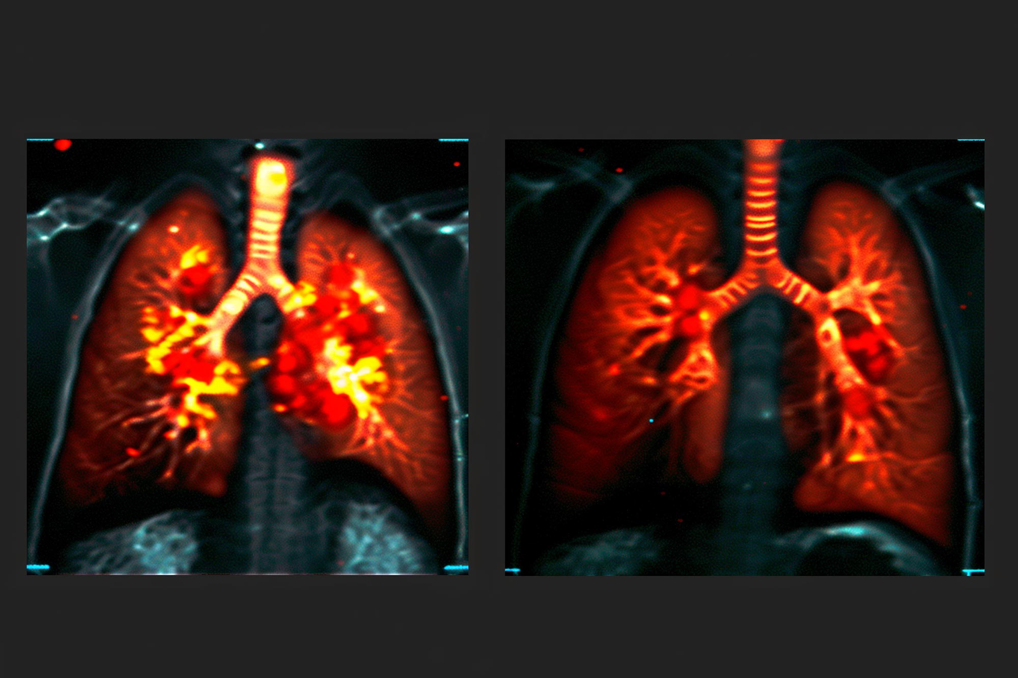
A novel lung scanning technique utilizing a safe gas visible on MRI scans provides groundbreaking insights into lung function, enabling real-time monitoring of respiratory treatments. Developed by researchers at Newcastle University, the method identifies poorly ventilated areas in the lungs, significantly enhancing the diagnosis and management of conditions like asthma, COPD, and complications in lung transplant patients. By inhaling perfluoropropane, a specialized gas, doctors can visualize air flow and pinpoint regions that improve with treatment.
The technique allows immediate assessment of treatment effectiveness, such as using bronchodilators like salbutamol, and facilitates early detection of lung function decline, vital for transplant recipients facing chronic lung dysfunction.
Clinical trials demonstrate its potential applications, giving healthcare providers an innovative tool for personalized patient care. The sensitivity of this imaging method may enable earlier intervention and better protective measures for transplanted lungs. Overall, this advancement in lung imaging could transform clinical management for various lung diseases, providing valuable data for individualized therapies and improving patient outcomes. The research is supported by the Medical Research Council and The Rosetrees Trust, highlighting its promise for future clinical applications.


